Overview
Project Title: 3D Model series – ‘Parapharyngeal Space, Neck Region and Floor of Mouth’
Project Sponsor: Mr Luke Reid, Lecturer in Anatomy
TILT Medical Artist: Eve Laws
3D models have proven to be extremely beneficial in student learning. Complex anatomical structures in this format aid and encourage information intake. 3D Models can be rotated, labelled, and explored in a manner that specimens cannot.
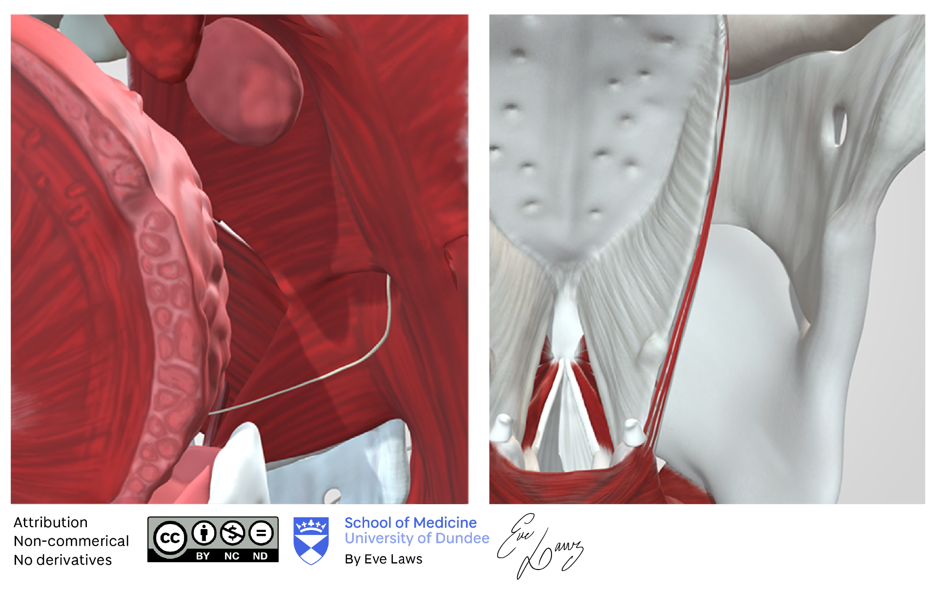
The models were created for ENT teaching and were aimed at the year 2 medical students. The three models would cover as follows – muscles and ligaments of the larynx, the pharynx and floor of mouth, and the parapharyngeal space. These were to be hosted on our TILT account at Sketchfab.com, all fully labelled.
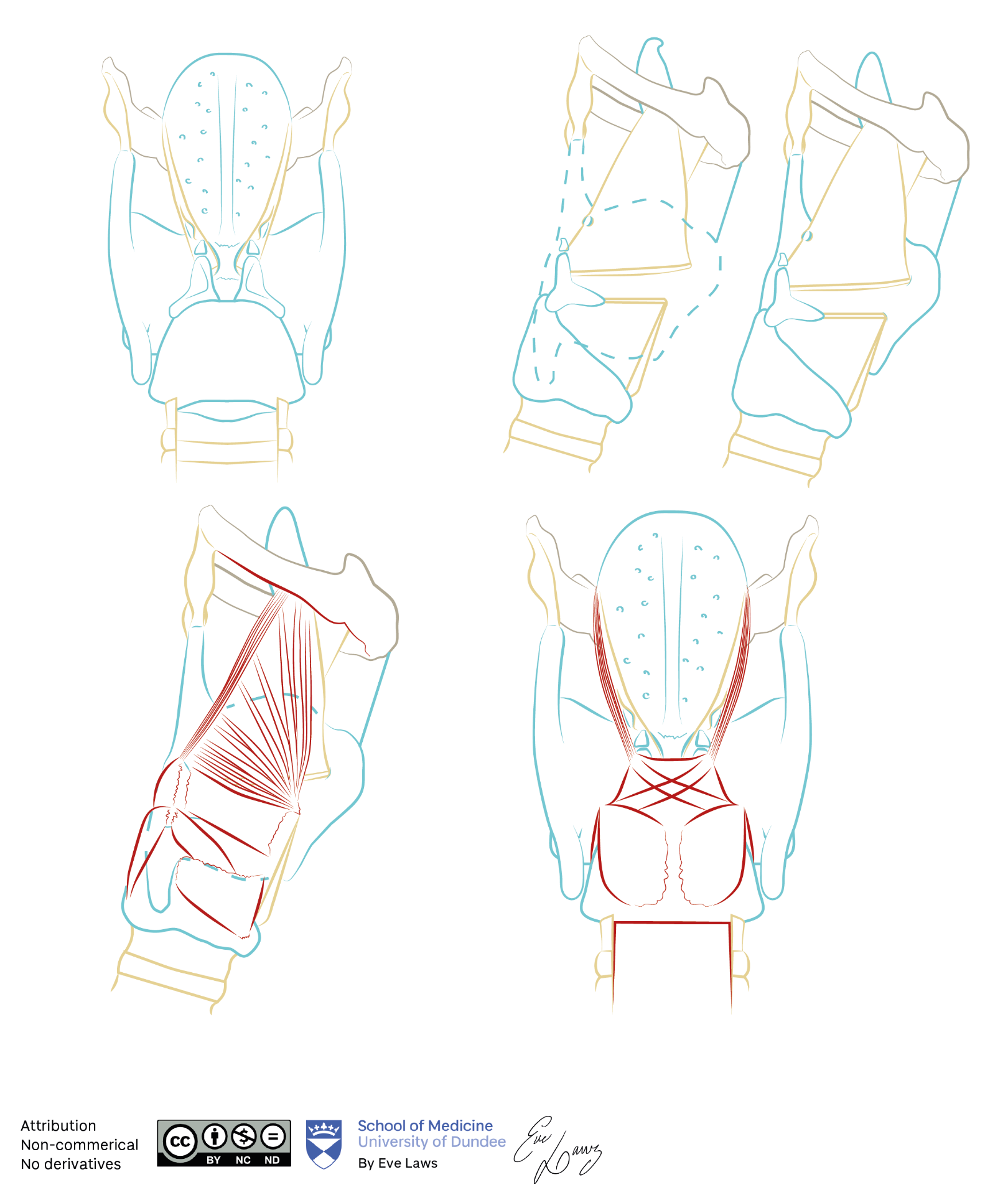
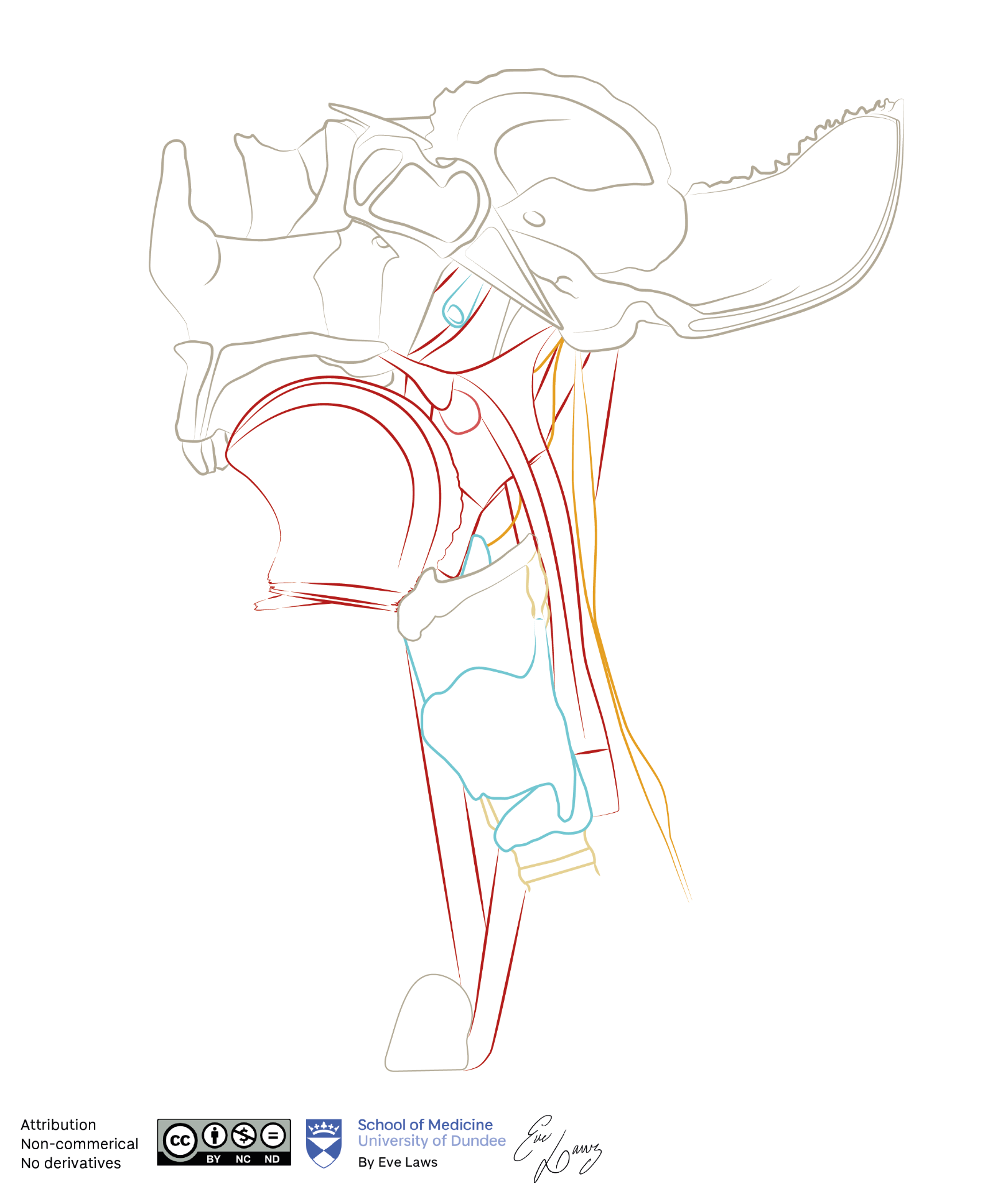
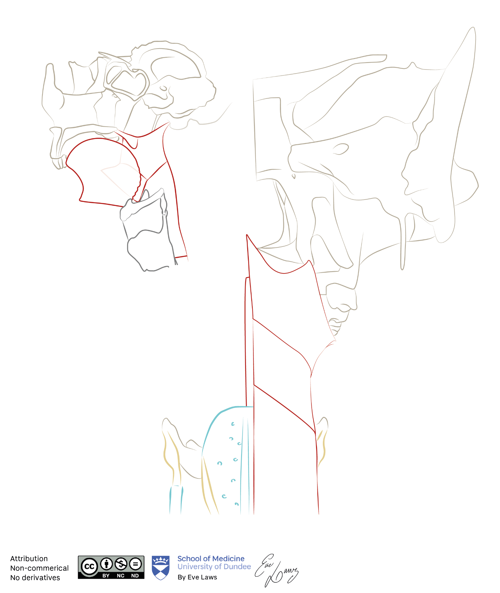
To complement the models, a series of illustrations for the students to colour and label were also created.

Illustrations focusing on the musculature and ligaments of the larynx


Linework Illustrations for the students to colour and label
