Overview
Project Title: ‘Olfactory Pathway’ Illustrations
Project Sponsor: Dr Richard Green, ENT Consultant
TILT Medical Artist: Eve Laws
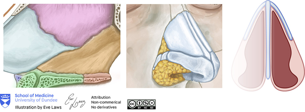
Detailed illustrations of the nasal/septal area were requested to aid in undergraduate teaching. One illustration required a sagittal view of the nasal area with key structures labelled (quadrangular cartilage, anterior, middle and posterior septal angle, the nasal spine, maxillary crest, the vomer, and perpendicular plate of the ethmoid bone).
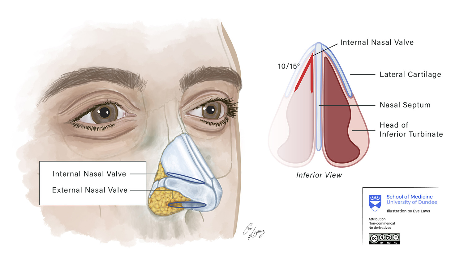
For the second illustration ,there would be a focus on the nasal cartilage (with a faint skin layer) at an anterolateral angle, with the internal and external nasal valve labelled. Alongside this would be a vector illustration (inferior view) with labels highlighting the lateral cartilage, nasal septum and head of the inferior turbinate labelled. As there were quite a few labels to add, it was important not to overcrowd the illustrations. For the skin layer, I experimented with a ‘painterly’ style of brushstrokes, adding a unique element to the illustration.

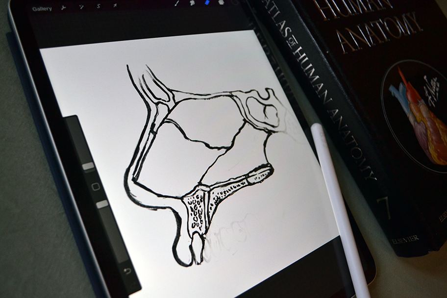
The drafting stage, created in Procreate on the iPad 2020
Final Products


In July of 2021, the Olfactory Pathway illustrations won Silver in the Institute of Medical Illustrators annual awards. The artwork now features on their website and has been published in the winners catalogue, with the Journal of Visual Communication in Medicine to follow.
