Overview
Project Reviewers: Dr. Penny Lockwood, Dr. David Stewart, Dr. Patrick Spielmann
Content Expert: Dr Patrick Spielmann
Medical Artist: Annie Campbell
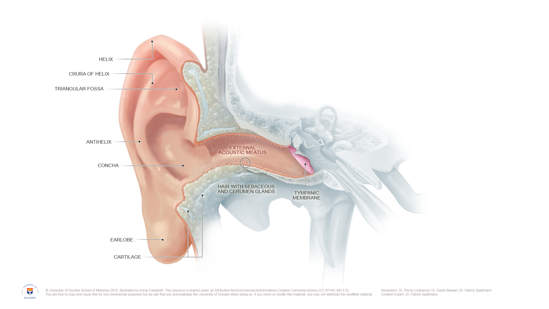
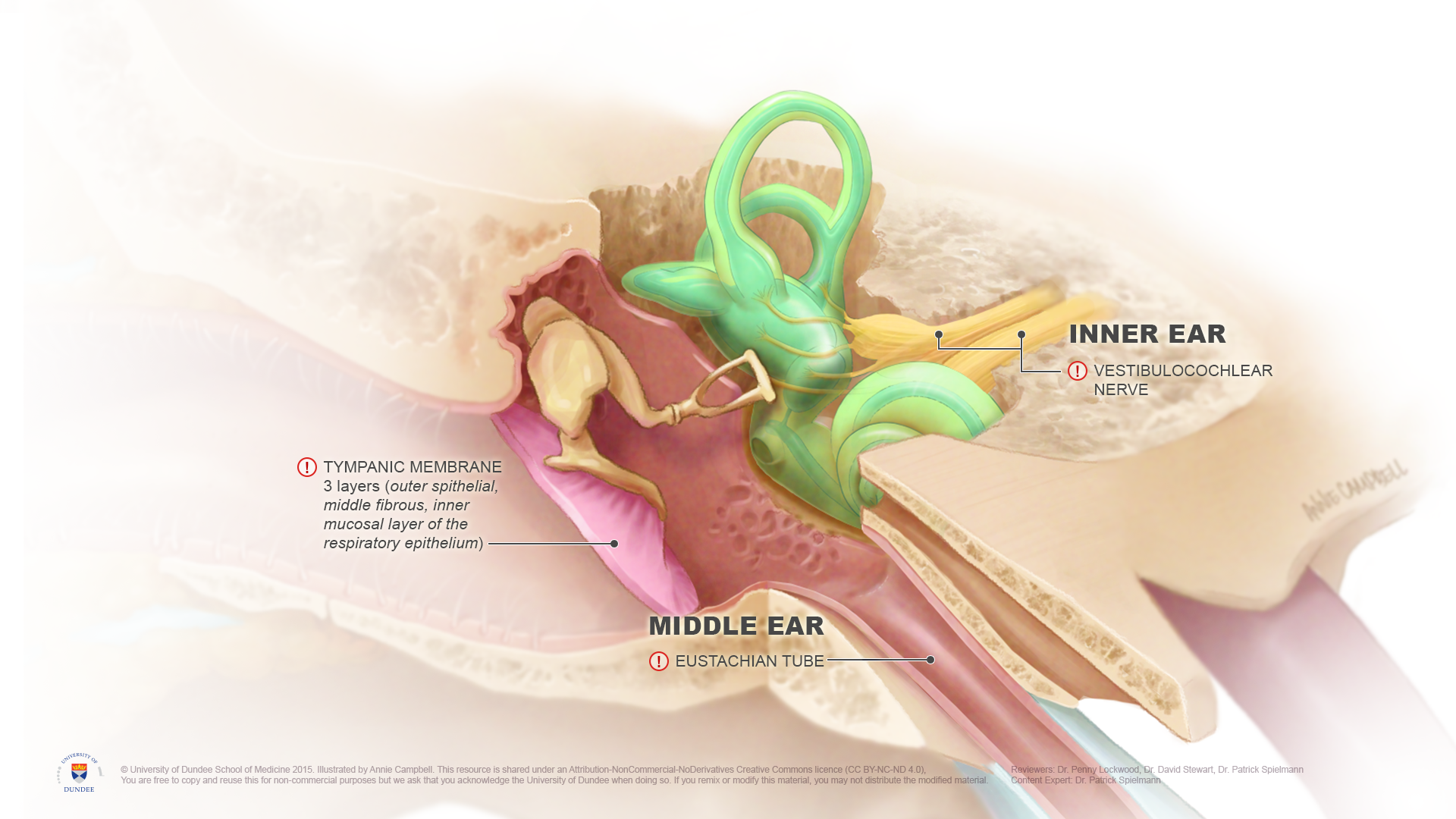
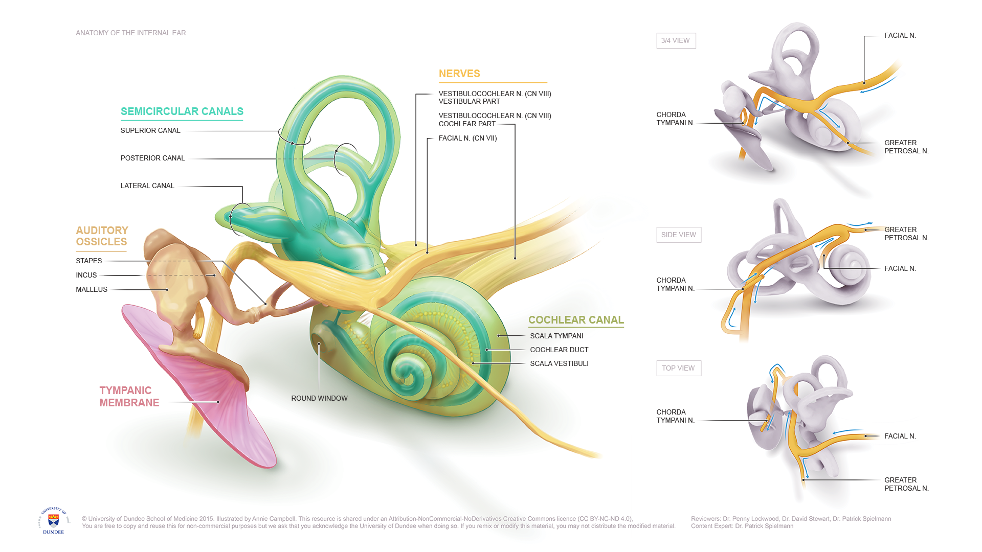
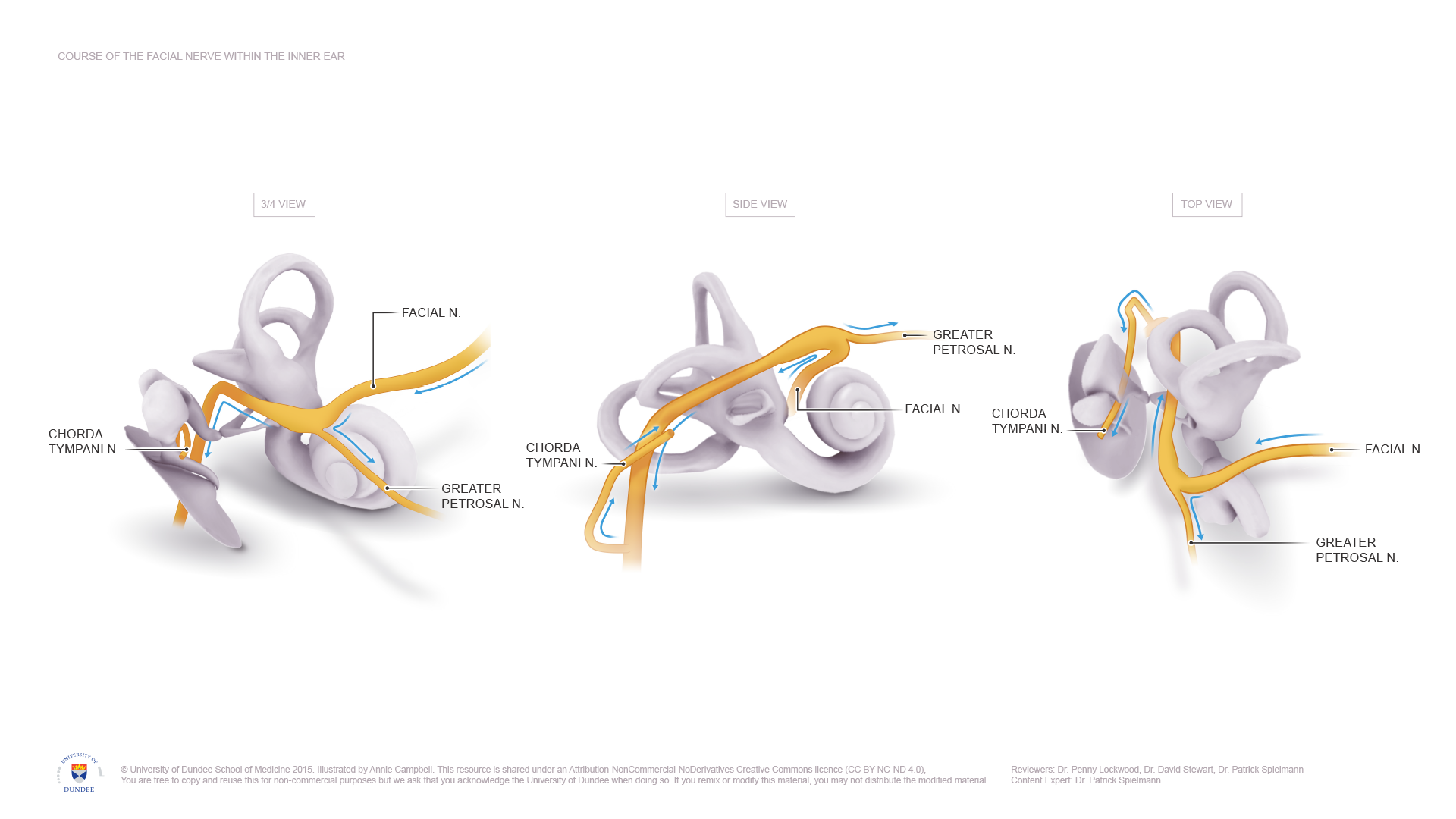
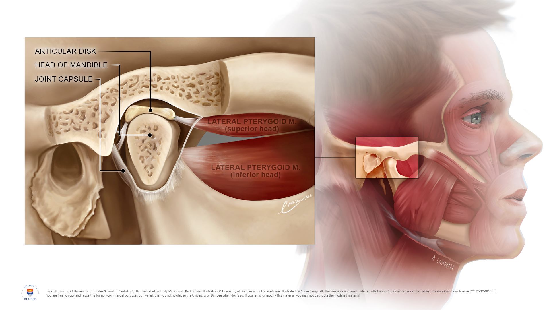
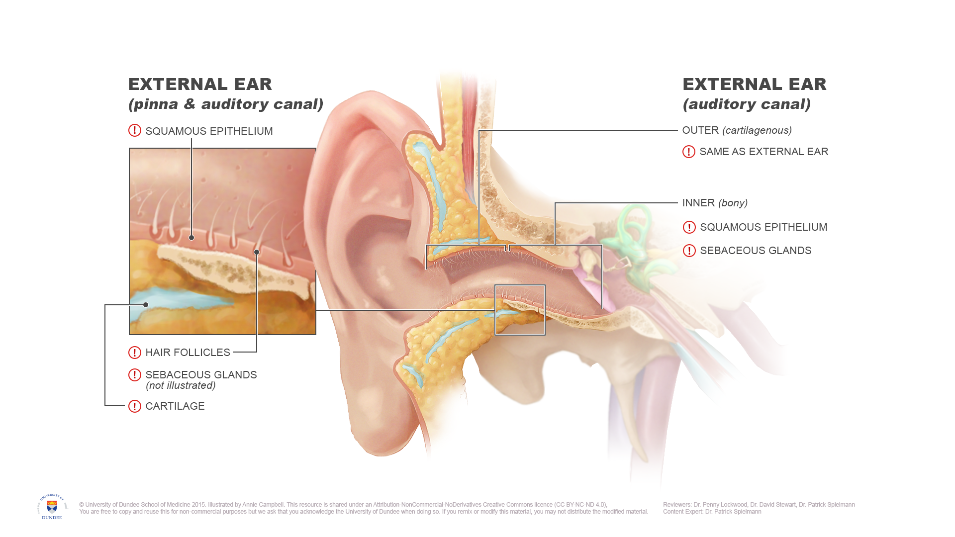
A series of illustrations was created for lecture material on the anatomy of earache. The illustrations would be used in group activity in identifying the possible causes and related structures. These illustrations would help students generate a differential diagnosis for causes of earache originating in the ear itself. Secondary structures potentially causing referred pain and their relation to the cranial nerves that supply them were also a specific focus. These secondary structures and their cranial nerves can be viewed in our animation collection here.
Final Products
